Try CBCT/TMJ Training online
Master the Art of Temporomandibular Disorder Diagnosis: A Cutting-Edge Learning Tool for Precision and Proficiency in Radiological Evaluation
Temporomandibular disorders (TMDs) exert multifaceted impacts on affected patients, manifesting as symptoms such as headaches, restricted jaw mobility, and audible joint sounds. These conditions afflict approximately 12% of the population, underscoring the significance of understanding and managing these disorders. The Diagnostic Criteria for TMD established by Dworkin and LeResche in 1992 has played a pivotal role in standardizing the identification of TMDs in patients. To assess osseous alterations within the temporomandibular joint, studies conducted by Ahmad et al. have advocated the use of advanced imaging techniques, such as computed tomography (CT) and, Cone Beam Computed Tomography (CBCT).
The emergence of CBCT signifies a paradigm shift in radiological evaluation, offering enhanced precision and three-dimensional visualization. In response to the growing need for expertise and proficiency in this evolving domain of radiological evaluation, this platform has been established. Its primary purpose is to foster the cultivation of practical skills and knowledge, thereby empowering practitioners and enthusiasts to navigate the complexities of TMD diagnosis and treatment through radiological methods. Through the dissemination of information and the encouragement of hands-on practice, this initiative strives to elevate the standards of care and promote a comprehensive understanding of temporomandibular disorders within the medical community.

Test
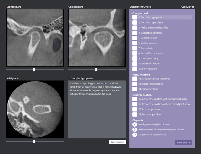
The examination comprises 15 cases of cone beam computed tomography (CBCT) scans of the temporomandibular joint (TMJ), with evaluations conducted in accordance with the Diagnostic Criteria for Temporomandibular Disorders (DC/TMD). A detailed explanation of each criterion can be obtained in conjunction with the analysis of each case, allowing for a comprehensive understanding of the diagnostic process.
Upon completion of the fifteenth case, the results are presented alongside evaluations conducted by three seasoned specialists in the field. This comparative analysis offers valuable insights into the accuracy and consistency of the assessments made. It is worth noting that participants are afforded the opportunity to undertake the test multiple times, enabling them to refine their skills and enhance their proficiency in diagnosing temporomandibular disorders based on CBCT images. This iterative approach not only facilitates learning and knowledge acquisition but also contributes to the ongoing development of expertise in the realm of TMJ evaluation.
The study about this learning tool
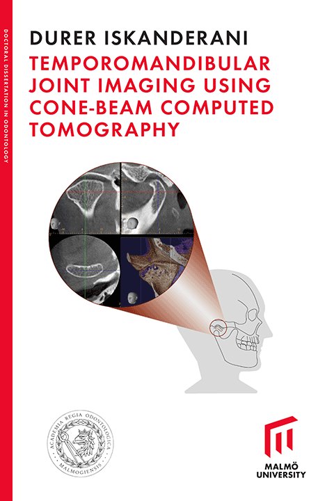
This learning tool is one of the projects in Dr. Iskanderani's PhD dissertation. This learning tool is one of the projects in Dr. Iskanderani's PhD dissertation. The research project delves into the development and efficacy of this innovative educational resource. Dr. Iskanderani's work sheds light on the intricate nuances of temporomandibular disorders diagnosis through radiological methods and elucidates the impact of this learning tool on the skills and knowledge acquisition of practitioners in the field.
For those interested in exploring the findings and insights gleaned from this study, the complete article is accessible to all readers. To access the detailed research and analysis conducted by Dr. Iskanderani, please click on the provided link below.
Warm regards,
Kristina Hellén-Halme
Professor and Head of Oral and Maxillofacial Radiology,
Faculty of Odontology, Malmö University
kristina.hellen-halme@mau.se
